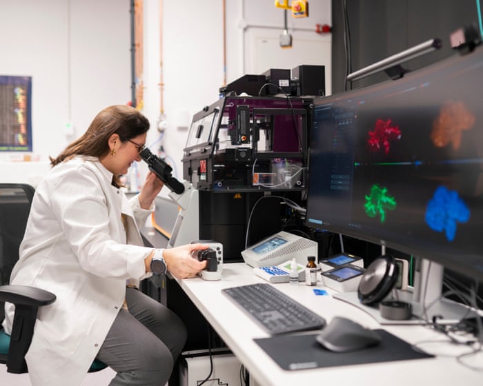
Researchers at the Rosalind Franklin Institute are using a high-resolution microscope to study the cellular mechanisms of inflammatory bowel disease.
Key facts
- •More than half a million people in the UK live with inflammatory bowel disease.
- •IBD is most commonly diagnosed in individuals between the ages of 15 and 40.
- •The new microscope is the only one of its kind in the UK, according to researchers.
- •The Rosalind Franklin Institute will receive £67 million in funding over five years beginning in April 2027.
- •The Science and Technology Facilities Council must achieve £162 million in savings by 2030.
Researchers at the Rosalind Franklin Institute in Oxfordshire have unveiled a specialized microscope, nicknamed "Curie," designed to study the cellular mechanisms of inflammatory bowel disease (IBD). The technology offers 10 times the resolution of conventional light microscopy, allowing scientists to observe biological structures as small as 20nm. Experts hope this increased clarity will help identify the root causes of IBD and lead to more targeted treatments for the condition.
By the numbers
Advanced Imaging Technology
The microscope utilizes stimulated emission depletion (STED) technology, a technique that earned a Nobel prize in 2014. By using two lasers, the device can isolate fluorescence to a very small region, allowing it to distinguish structures that are otherwise too close together to be seen by standard equipment. The system also features deformable mirrors to correct optical distortions and a temperature-controlled stage, which enables the study of living cells and mini-organs at near-body temperatures.
Research into Cellular Barriers
PhD student Dimitrios Ioannidis is currently using the microscope to examine how cells lining internal organs, such as the intestines, form protective barriers. By observing protein complexes from fetal development through adulthood, researchers aim to identify structural changes that may contribute to disease. Comparing healthy tissue to diseased tissue could potentially reveal the root causes of conditions like Crohn’s disease and ulcerative colitis.
Funding and Institutional Context
The unveiling of the microscope coincided with the announcement of £67 million in funding for the Rosalind Franklin Institute over five years starting in April 2027. This funding is provided by the Engineering and Physical Sciences Research Council. The announcement occurs amid broader budgetary challenges for UK science facilities, as the Science and Technology Facilities Council faces a requirement to make £162 million in savings by 2030 due to rising costs.
Advertisement
This article was independently rewritten by ManyPress editorial AI from reporting originally published by The Guardian Health.